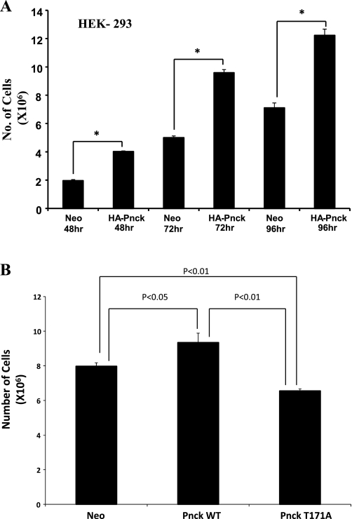
Fig. 6.
Pnck induces cellular proliferation in HEK 293 cells. A: enhanced proliferation of Pnck-overexpressing HEK-293 cells. Nine 60-mm dishes were plated with equal numbers of Neo and HA-Pnck cells in complete medium, and a set of triplicate dishes of each cell lines were trypsinized at 48, 72, and 96 h and counted by hemocytometer. A commercial statistical software package (GraphPad Prism, version 4.03) was used to analyze data, which are expressed as means ± SD of triplicate samples. One-way analysis of variance followed by Turkey's multiple-comparisons tests were used to determine the statistical significance of differences between groups. *P < 0.001, statistically significant difference between the compared groups connected with solid lines. B: mutant (T171A) Pnck is unable to induce cellular proliferation. Neo, HA-Pnck, and HA-Pnck-T171A HEK-293 cells were plated (0.5 × 10⁶ per dish) in triplicate in 10% heat-inactivated serum containing DMEM and counted after 96 h by hemocytometer. A representative example of three experiments is presented. A P < 0.05 value was considered significant.

