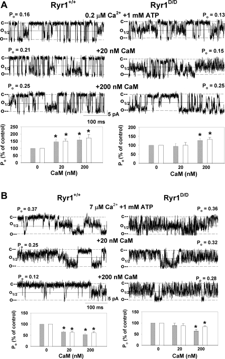
Fig. 2.
Effects of CaM on single Ryr1+/+ and Ryr1D/D channel activities. Membranes isolated from skeletal muscle of Ryr1+/+ and Ryr1D/D mice were fused with lipid bilayer as described in experimental procedures. Representative single-channel currents (downward deflections from closed levels, c–) were recorded at −35 mV at 0.2 μM (A) and 7 μM (B) cis, cytosolic free Ca2+ in the presence of 1 mM ATP as described in experimental procedures in the absence of CaM (top traces) and following the addition of 20 nM (middle traces) and 200 nM (bottom traces) cis CaM. Channel open probabilities (Po) obtained from 2-min recordings in the absence of CaM were 0.15 ± 0.02 (n = 21) and 0.14 ± 0.02 (n = 29) at 50% threshold setting, 0.30 ± 0.04 and 0.23 ± 0.02 at 25% threshold setting at 0.2 μM Ca2+ and 1 mM ATP, and 0.27 ± 0.05 (n = 14) and 0.26 ± 0.05 (n = 11) at 50% threshold setting and 0.38 ± 0.05 and 0.48 ± 0.05 at 25% threshold setting at 7 μM Ca2+ and 1 mM ATP for Ryr1+/+ and Ryr1D/D, respectively. Normalized Po values (bottom) were obtained from 2-min recordings by setting the threshold level at 25% (open bars) and 50% (gray bars) of the current amplitude between the closed (c) and open (o) channel states. Data are means ± SE of 9 and 15 (A) and 14 and 11 (B) channel recordings for Ryr1+/+ and Ryr1D/D, respectively. *P < 0.05 compared with respective control (−CaM).

