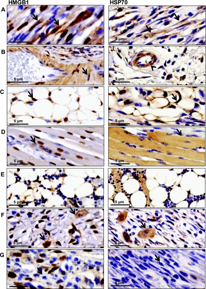
Fig. 4.
Endogenous danger signal expression is induced in cells native to injured tissue. Representative high-power views (×100) of immunohistochemical staining for HMGB1 (left) or HSP70 (right) obtained 3–5 mm distal to tail wounds at 3 wk postoperatively. Positive cellular staining for both markers was present in fibroblasts (arrow) (A), vascular endothelium (arrowhead) (B) including smooth muscle cells of vascular endothelium (arrow), and adipocytes (C) (arrow). Skeletal muscles cells in close proximity to the wound stained positively for HMGB1 but not HSP70 (D). Bone marrow (E) and macrophages (F) stained positively for HSP70 and HMGB1 at postoperative week 3. Infiltrating lymphocytes and neutrophils remained negative (G). N = 5 slides were analyzed per group by a blinded pathologist and independent reviewer.

