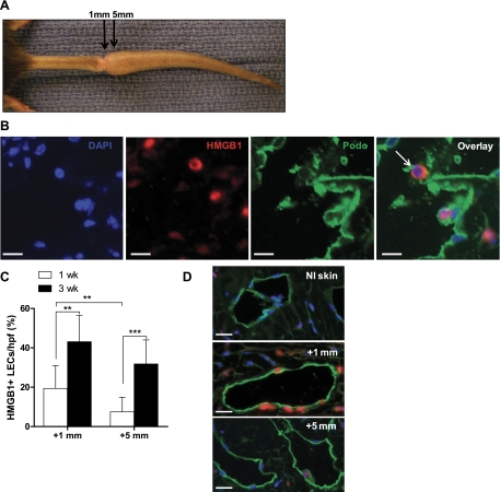
Fig. 5.
Tissue injury induces endogenous danger signal expression by lymphatic endothelial cells. A: gross representation of the injured mouse tail at 3 wks postoperatively; double immunofluorescence for HMGB1 and podoplanin was performed on longitudinal sections and analyzed immediately distal (within 1 mm) to the wound or 5 mm distal to the wound as indicated. B: DAPI (blue), HMGB1 [tetramethyl rhodamine isothiocyanate (TRITC), red], and podoplanin [fluorescein isothiocyanate (FITC), green] single stains and image overlays are displayed. Arrows indicate HMGB1-podoplanin double-positive cells. C: counts of positively stained cells were performed at 1 or 5 mm distal to the center of the wound. Mean percentage of HMGB1+ LECs per hpf is displayed. Bars represent means ± SD, **P < 0.01, ***P < 0.001. D: representative lymphatic vessel staining for HMGB1 within normal skin, 1 mm distal to tail wounds, or 5 mm distal to the wound.

