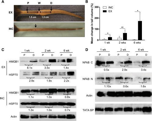
Fig. 6.
Endogenous danger signal expression is upregulated in sustained lymphatic fluid stasis. A: representative gross images of tails 3 wk postoperatively following tail skin excision and lymphatic disruption (EX, top) or circumferential incision without lymphatic disruption (INC, bottom). Protein from tail tissues was harvested 1.5 cm proximal (P) or distal (D) to the wound (W). B: mean percentage change in tail volume at 1, 2, and 6 wk postoperatively (from preoperative baseline) are displayed; bars represent means ± SD. *P < 0.05, **P < 0.01. C: Western blot analysis of pooled protein samples (n = 5) from the indicated locations (P, D) for HMGB1, HSP70, or actin at 1, 2, and 6 wk performed for both EX and INC groups. Protein from 1, 2, and 6 wk time points were run simultaneously. ImageJ analysis comparing band density at P and D and normalized to actin are displayed as fold change below blots. D: cytoplasmic and nuclear protein fractions isolated from the same tissue regions were subjected to Western blot analysis for NF-κβ (NF-κβ C and NF-κβ N, respectively), actin, or TATA BP nuclear loading control at 1, 2, and 6 wk. ImageJ analysis demonstrating fold change from P to D and normalized to loading control is displayed below blots. All blots were run in at least triplicate and fold change in band density represents an average of at least 3 ImageJ analyses.

