Fig. 8.
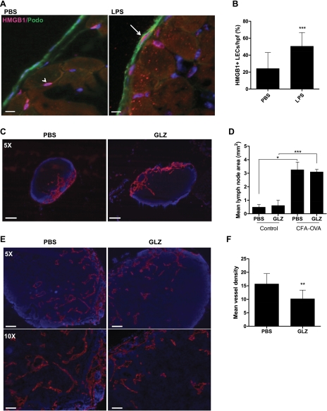
HMGB1 blockade attenuates inflammatory lymphangiogenesis in draining lymph nodes. A: HMGB1 (pink) and podoplanin (green) double immunofluorescence of mouse diaphragms (n = 6 per group) after 2 wk daily administration of PBS or LPS. Arrow indicates costained LEC. Note presence of positively stained cells in PBS-treated animals not colocalized with podoplanin (arrowhead). Scale bars represent 10 μm. B: HMGB1/podoplanin double-positive cells on the peritoneal surface of the diaphragm. Bars represent means ± SD, **P < 0.01, ***P < 0.001. C: lymphatic vessel endothelial hylaluronan (LYVE)-1 stains of popliteal nodes from animals treated with PBS or glycyrrhizin (GLZ). Scale bars represent 100 μm. D: mean lymph node area (mm2) for control or complete freund's adjuvant (CFA)-ovalbumin (OVA)-treated animals treated with PBS or GLZ (n = 6). Bars represent means ± SD, *P < 0.05, ***P < 0.001. E: LYVE-1 staining in lymph nodes harvested from animals treated with CFA-OVA and PBS (left) or GLZ (right). Scale bars represent 200 μm (top) and 100 μm (bottom). F: lymphatic vessel density in animals treated with CFA-OVA and PBS or GLZ. Bars represent means ± SD, **P < 0.01.

