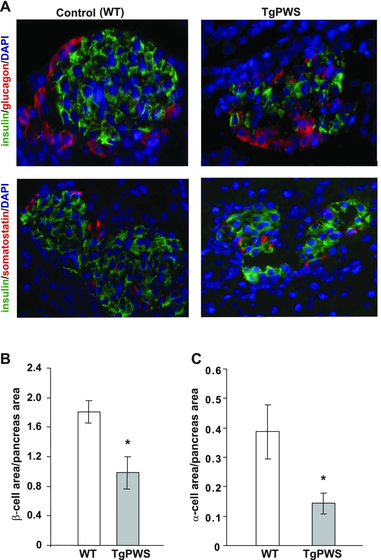
Fig. 4.
Altered islet morphology with reduced number of β- and α-cells in TgPWS pancreas compared with controls at P1. A: islets from TgPWS and WT pancreata were stained using antibodies against insulin (green) and glucagon or somatostatin (red) to detect β-, α-, and δ-cells, respectively. Cell nuclei were stained with 4,6-diamidino-2-phenylindole (DAPI; blue). B and C: β- and α-cell mass, respectively, quantified in pancreata from TgPWS and WT mice. Values represent percentage ± SE of β- and α-cell area normalized to the total tissue area of counted sections (n = 11 or 10 for TgPWS; n = 8 or 7 for WT for β- and α-cell area, respectively). *P < 0.05, significant differences between TgPWS and WT mice (independent t-test).

