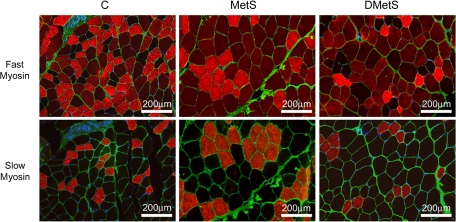
Fig. 2.
Immunostaining of plantaris (PLN) muscles of control, MetS, and DMetS diets with antibodies against fast and slow myosins. Fast 2a muscle fibers were identified by bright red staining, 2b/x by dull red staining, and type 1 by no staining (black) with antibody against fast myosin. Slow type 1 muscle fibers were identified by bright red staining with antibody against slow myosin. Anti-laminin antibody (green) was used for visualization of the muscle fiber boundaries.

