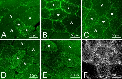
Fig. 6.
Staining of the IMCL droplets with BODIPY 493/503 in PLN muscle of the Ossabaw swine on control (A), MetS (B), and DMetS (C-F) diets. Type 1 and type 2a muscle fibers (*) accumulated a much higher amount of IMCL droplets than type 2b/x fibers (^). Some of the type 1 and 2a muscle fibers had very high accumulation of the IMCL droplets (D). Tangential sections (E) and three-dimensional reconstruction (F) showed that IMCL droplets were arranged in continuous rows.

