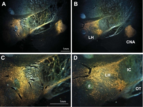
Fig. 1.
Darkfield photomicrographs of coronal sections showing wheat germ agglutinin-horseradish peroxidase (WGA-HRP) reaction product in the ventral forebrain after an infusion of the tracer into the pontine parabrachial nuclei. Dorsal is up, medial to the left. Top: reaction product (orange) is visible in the lateral hypothalamus (LH) and the central nucleus of the amygdala (CNA). A: LH lesion. B: control. Note that the CNA is filled with reaction product in both brains. Bottom: higher power photomicrographs of the LH reveal a pattern of dots and haze. The dots represent retrogradely labeled cell bodies; the haze is a mixture of retrograde and anterogradely labeled neural processes, i.e., axons and preterminal arbors. C: LH lesion. D: control. The core of the LH lesion eliminated all retrograde and anterograde labels. More medially, in the perifornical region, considerable reaction product remains. F, fornix; IC, internal capsule; OT, optic tract.

