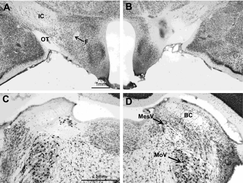
Fig. 4.
Photomicrographs of coronal sections through the LH (top) and the parabrachial nuclei (PBN; bottom) in a rat (no. 06–17) with asymmetric lesions (NeuN stain). A: intact left LH. B: lesioned right LH. C: lesioned left PBN. D: intact right PBN. The acellular area in C extends into the supratrigeminal area above the motor trigeminal nucleus (MoV) and the locus coeruleus medial to the mesencephalic trigeminal nucleus (MesV). BC, brachium conjunctivum. Magnification in C and D is double that of A and B.

