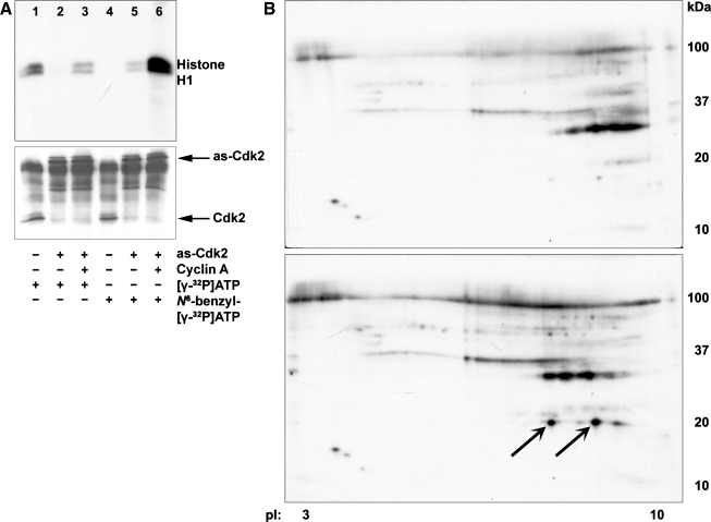
Fig. 2.
A: activities of Cdk2s using ATP analogs. The activity of wild-type Cdk2 and analog-sensitive Cdk2 (as-Cdk2) was determined by histone H1 kinase assay using [γ-32P]ATP (lanes 1–3) or N6-benzyl-[γ-32P]ATP (lanes 4–6). Cells were either untreated (lanes 1, 4), transduced with as-Cdk2mCherry (lanes 2, 5), or transduced with as-Cdk2mCherry plus cyclin A adenoviruses (lanes 3, 6). Wild-type Cdk2 was immunoprecipitated by Cdk2 antibody (lanes 1, 4). The as-Cdk2 was immunoprecipitated using Discosoma red fluorescent protein (DsRed; anti-mCherry) antibody (lanes 2, 3, 5, 6). An immunoblot using Cdk2 antibody showed that similar amounts of Cdk2 were immunoprecipitated either with anti-Cdk2 (bottom, lanes 1, 4) or anti-DsRed (bottom, lanes 2, 3, 5, 6). B: 2-dimensional (2D) gel electrophoresis. Cdk2-bound TKPTS cellular proteins were immunoprecipitated from untreated cells (top) or cells exposed to cisplatin for 24 h (bottom). Proteins were separated using 2D electrophoresis. Proteins were identified as p21 labeled with arrows.

