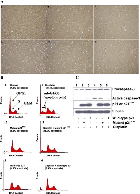
Fig. 4.
A: apoptosis assay by light microscopy. TKPTS cells were untreated (panel 1) and exposed to 25 μM cisplatin for 24 h (panels 4–6). Cultures were transduced with adenovirus-expressing mutant p21S78D (panels 2, 5) and wild-type p21 (panels 3, 6) for 48 h. B: apoptosis assay by fluorescence-activated cell sorter (FACS) analysis. Cells were treated the same as described in A. Cells were stained with propidium iodide (PI) and analyzed using FACSCalibur. The cells were grouped into sub-G1/G0, G1/G0, S, and G2/M phases using a cell cycle analysis program (WinMDI 2.8). Cells in sub-G1/G0 were considered apoptotic. C: mutant p21S78D lost its ability to prevent caspase cleavage induced by cisplatin. Source of protein extracts as in A. Both full-length (procaspase) and cleaved (active) caspase-3 are indicated. Expression of wild-type and mutant p21 was detected by immunoblot using p21 (F5) antibody. α-Tubulin was used as a loading control.

