Fig. 8.
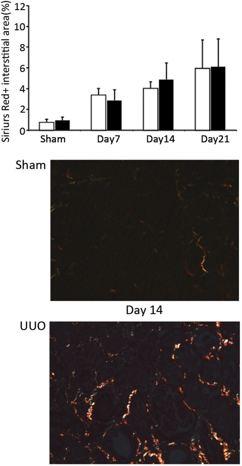
Interstitial fibrosis is similar in Vtn+/+ and Vtn−/− mice after UUO. The area of the interstitium occupied by picrosirius red-positive interstitial collagen fibrosis quantified by computer-assisted image analysis of stained kidney sections progressively increased with time after UUO. Differences between the Vtn+/+ and Vtn−/− mice (n = 5–12/group) were not significantly significant on days 7, 14, and 21. Top: bar graph represents results expressed as a mean positive interstitial area ± 1 SD. Bottom: representative photomicrographs of tissue sections illustrate the increase in sirius red-positive interstitial collagen deposits 14 days after UUO surgery compared with sham surgery. Magnification ×200.

