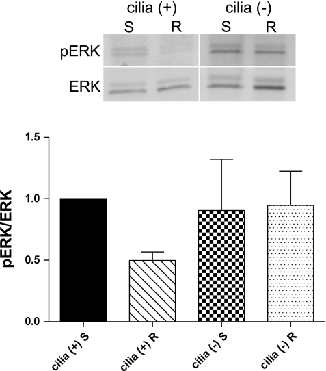
Fig. 3.
Western blot analysis for phosphorylated ERK (pERK) in cilia (+) and cilia (−) cells at 12 h. Cells were incubated while stationary (S) or rotated (R) at 1 Hz to stimulate cilia bending. pERK levels were compared with total ERK levels, and relative band density is presented compared with stationary cilia (+) cells. Although not significant by 1-way ANOVA, there was significance between stationary and rotating conditions in cilia (+) cells when examined by t-test; n = 5.

