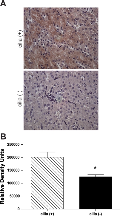
Fig. 6.
Immunohistochemical analysis of RKIP in mouse kidney sections. A: kidney sections from cilia (+) and cilia (−) mice were stained for RKIP and imaged at ×40 magnification in a random order. B: densitometry analysis determined the relative density of staining. *P < 0.05; n = 3 [cilia (+)] and n = 4 [cilia (−)], 1 slide/mouse, 10 fields/slide.

