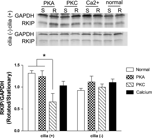
Fig. 7.
Western blot analysis for RKIP in cilia (+) and cilia (−) cells following incubation with inhibitors for PKA, PKC, or a calcium chelator for 12 h. Cells were incubated while stationary (S) or rotated (R) at 1 Hz immediately after addition of treatment. RKIP levels were normalized to GAPDH, and relative band density is presented as amount in rotated cells compared with amount in stationary cells. *P < 0.05; n = 3.

