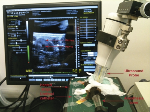
Fig. 1.
Mouse undergoing live cystogram. Representative image showing a mouse with its nose in the anesthesia cone and on a heated table that can monitor breathing and heart rate. PE-10 tubing used to catheterize the mice is visible as is the ultrasound probe that is pressed against the shaved flank. The ultrasound image of the kidney is seen on the monitor.

