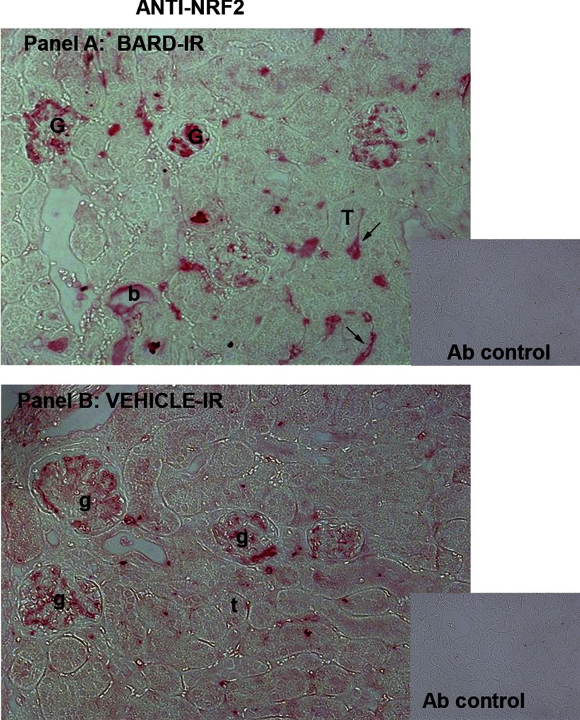
Fig. 9.
Increased Nrf2 in BARD- vs. vehicle-treated ischemic renal cortex. A: anti-Nrf2 immunoperoxidase staining of BARD-treated ischemic kidneys at 8-h reperfusion. G, intensely Nrf2-positive glomeruli; T, one of many Nrf2-negative tubules; b, one rare Nrf2-positive tubule. Black arrows indicate peritubular Nrf2-positive “capillaries.” Capillaries are provisionally identified in this figure, and definitively identified in Figs. 11 and 12 that show double staining of CD31 and Nrf2. Inset: control antibody. B: anti-Nrf2 immunoperoxidase in vehicle-treated ischemic kidneys at 8-h reperfusion. Glomeruli that are less intensely stained than in the BARD-treated kidney in A are indicated as g, and t indicates one of many tubules. Inset: control antibody.

