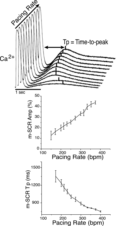
Fig. 2.
Shown at the top are superimposed intracellular calcium traces measured from a single recording site during the termination of pacing at increasing rates. As pacing rate increases (400–160 beats/min), multicellular spontaneous calcium release (m-SCR) amplitude increases and the time-to-peak (Tp) occurs earlier. Summary data show that faster pacing rates increase m-SCR amplitude (bottom left) and decrease m-SCR Tp (bottom right).

