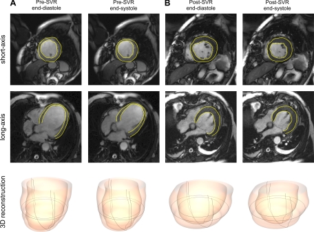
Fig. 1.
Magnetic resonance image showing a short-axis slice (top) at the level of the equator and long-axis slice (middle) at end diastole and end systole, with the left ventricular endocardial and epicardial surfaces denoted by yellow lines. Its corresponding three-dimensional (3-D) reconstructed left ventricular shape (bottom) at end diastole and end systole are shown before (A) and after (B) surgical ventricular restoration (SVR).

