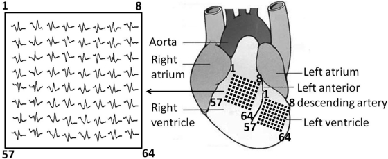
Fig. 1.
Diagram showing the multielectrode array placed on the surface of the left ventricle (LV) or right ventricle (RV), with channel 1 near the base of the heart and channel 64 near the apex. Position of the array was determined using the anatomical landmarks of the left anterior descending artery, the aorta, and the atria. Inset: typical set of electrogram traces.

