Fig. 2.
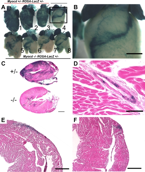
Myocardin−/− ESCs readily form atrial ventricular myocytes and coronary smooth muscle cells but not ventricular myocytes. Hearts from chimeric animals at 10 days of age were dissected out and stained with X-gal. A: myocardin−/−/LacZ+ (Myocd−/− ROSA-LacZ+/−) ESCs failed to contribute to the ventricles (hearts 4–8) compared with myocardin+/−/LacZ+ (Myocd+/− ROSA-LacZ+/−) cells (hearts 1–3). Scale bar, 1 mm. The myocardin−/−/LacZ+ cells typically formed a superficial band of myocytes extending from the aortic root in the left ventricle, across the ventricular septum, and continuing in a band across the right ventricle. B: higher magnification of heart 4 (box in A) shows myocardin−/−/LacZ+ cells contributed to coronary vasculature (see also enlarged image in D) and atria. C: longitudinal sections show abundant myocardin+/−/LacZ+ cells contributing to the ventricles (top) compared with myocardin−/−/LacZ+ cells (bottom). D: high magnification of a coronary artery from a myocardin−/− chimera showing abundant myocardin−/−/LacZ+ cells in a coronary artery. Scale bar, 100 μm. E and F: saggital sections of the right ventricle show that myocardin−/−/LacZ+ cells contributed to a superficial strip across the anterior right ventricle in an inferior (E) and superior section (F) across the heart. Scale bar, 500 μm. Results were consistent over 30 myocardin−/−/LacZ+ animals at ages from 10 days to 6 mo.

