Fig. 3.
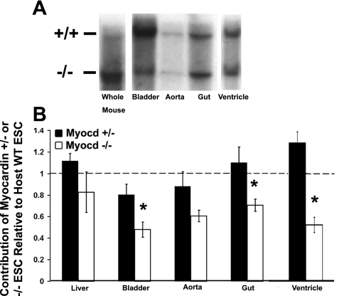
Quantitative Southern analyses show reduced investment of myocardin−/− cells in visceral SMC tissues, bladder, and cardiac ventricles. Bladder, aorta, small intestine/gut, and ventricle were dissected from 28-day-old myocardin+/−/LacZ+ control and myocardin−/−/LacZ+ animals, lysed, DNA purified and electrophoresed, and transferred to a nylon membrane for Southern analysis. A: representative Southern blots. If cells contributed equally to all tissues, the ratio between the +/+ lane, representing cells derived from ESCs from the host blastocysts, which have myocardin wild-type (WT) alleles, and the −/− lane, representing cells derived from ESCs containing knockout alleles, would be equivalent in all tissues (i.e., a ratio of 1). The “whole mouse” lysate lane represents homogenized tissue from an entire mouse (less dissected organs and tissues), and the ratio between the bands in this lane represents the overall contribution of chimeric cells in the individual animal assayed (i.e., %chimerism). Analysis of tissues from myocardin−/−/LacZ+ chimeras showed a decreased ratio of cells derived from ESCs with myocardin−/− alleles in visceral SMC tissues and ventricles compared with that observed in whole mouse lysates, suggesting myocardin is required for appropriate contribution of ESC-derived cells to these tissues. B: results of densitometric analysis of multiple independent Southern blots. Myocardin−/−/LacZ+ chimeras showed a statistically significant decrease in the contribution of myocardin−/− ESCs in the bladder, gut, and ventricles compared with WT ESCs from the host blastocysts (n = 4 myocardin+/−/LacZ+ and 5 myocardin−/−/LacZ+). Values are means ± SD. *P < 0.05. Heterozygous myocardin+/− ESCs exhibited no significant change in their contribution to any of these tissues, indicating that loss of 1 myocardin allele had no effect.

