Fig. 4.
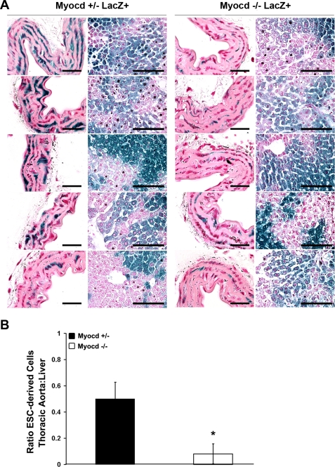
Histological analysis shows reduced investment of myocardin−/− ESCs into vascular SMC lineages. Tissues were fixed and stained for LacZ. Aortic strips were prepared en face and sectioned, and LacZ+ and LacZ− nuclei were counted to determine the percentage of cells that were ESC derived in each tissue. A: cross sections of aortas prepared en face and examined with nuclear fast red (NFR) staining (left) at high magnification show fewer LacZ+ cells in myocardin−/−/LacZ+ aortas compared with liver tissue (right) relative to myocardin+/−/LacZ+ controls. Aorta: scale bar, 25 μm; liver: scale bar, 100 μm. B: quantification of the ratio of LacZ+ nuclei in the thoracic aorta relative to the liver shows a markedly decreased contribution of myocardin−/−/LacZ+ ESCs to vascular SMCs compared with the contribution of myocardin+/−/LacZ+ ESCs. Values are means ± SD. *P < 0.05.

