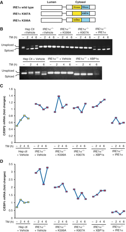
Figure 6.
Overexpression of IRE1α suppresses upregulation of CEBPβ and CEBPδ in Ire1α-null hepatocytes. (A) Depiction of domain structures for wild-type and mutant versions of IRE1α protein. K599A, IRE1α kinase mutant; K907A, IRE1α RNase mutant. Ire1α-null or control hepatocytes were infected with recombinant adenoviruses expressing different versions of IRE1α, spliced Xbp1 mRNA, or empty vector control. The infected hepatocytes were then treated with TM (2 μg/ml) for the times indicated. (B) Semiquantitative RT–PCR analysis of spliced and unspliced Xbp1 mRNAs in the Ire1α-null (Ire1αfe/−CRE) or control (Ire1αfe/fe) hepatocytes. (C, D) Quantitative real-time RT–PCR analysis of C/ebpβ and C/ebpδ mRNAs in adenovirus-infected hepatocytes. At 48 h after infection of Ire1α-null or control hepatocytes with the indicated adenoviruses, the hepatocytes were treated with TM (5 μg/ml) for the times indicated. Expression values of C/ebpβ and C/ebpδ mRNAs were normalized to β-actin mRNA. Fold changes of mRNA were measured by comparing to the expression level of mRNA in one of the empty virus-transfected control cells. Hep ctl, control (Ire1αfe/fe) hepatocytes; Ire1α−/−, Ire1α-null (Ire1αfe/−CRE) hepatocytes. A full-colour version of this figure is available at The EMBO Journal Online.

