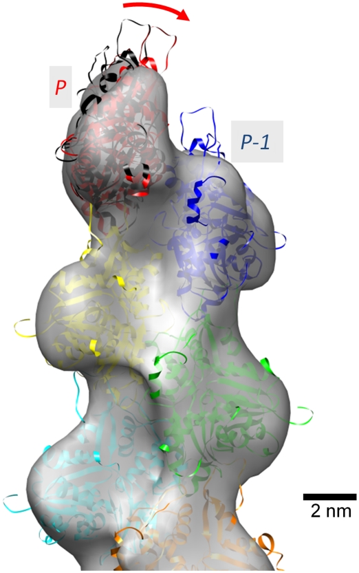
Figure 3.
The subunit P tilted towards the subunit P-1. Superposed on the EM map of Figure 1 are ribbon models of subunits (Oda et al, 2009), each coloured separately, which were fitted to the EM map without energy minimization and MD. The fitted subunit P to the EM map (red) tilts towards subunit P-1 by ∼12° compared with the subunit according to the canonical actin helical symmetry (black). Substantial parts of the DNase I binding loops of subunits P and P-1 protrude from the envelope, indicating that these loops might adopt different conformations at the pointed end compared with those within the filament.

