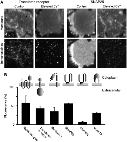
Figure 1.
Rising intracellular Ca2+ dramatically diminishes membrane protein immunostaining intensities. (A, B) PC12 cells were treated for 5 min with 20 μM ionomycin in the absence or presence of extracellular Ca2+, followed by the generation of membrane sheets and immunostaining for a variety of structurally diverse membrane proteins. (A) (Upper panels) TMA-DPH staining for the visualization of phospholipid membranes, indicating location and integrity of the basal plasma membranes; (lower panels) immunostaining for transferrin receptor (left) or SNAP25 (right). (B) Quantification of Ca2+-dependent decreases in immunostaining intensity. Remaining fluorescence after Ca2+ treatment was normalized to the values obtained in the absence of Ca2+ and expressed as percentage. Values are means±s.e.m. (n=3 independent experiments; 31–78 membrane sheets were analysed for each condition in one experiment).

