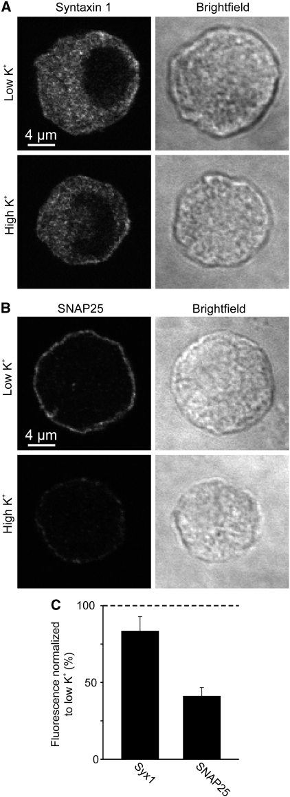
Figure 5.
Membrane protein redistribution after depolarization-induced calcium channel opening. (A, B) Bovine chromaffin cells were treated for 30 s with low or high potassium Ringer solution at 37°C, fixed and immunostained for syntaxin (A) or SNAP25 (B). Confocal micrographs from equatorial sections in the immunofluorescence (left) and in the brightfield (right) channels are shown. Depolarization-induced decrease of plasmalemmal immunofluorescence was analysed by linescans placed at the periphery of the cell (for details see Materials and methods). (C) High potassium values were related to corresponding low potassium values (set to 100%). Values are means±s.e.m. (n=4 independent experiments).

