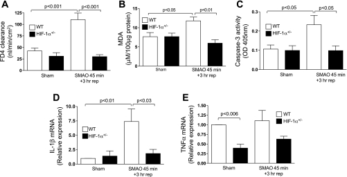
Fig. 2.
HIF-1 is involved in gut barrier dysfunction and the mucosal inflammatory response. WT and HIF-1α+/− mice were subjected to SMAO or sham for 45 min and 3 h of reperfusion. A: ex vivo everted gut sac permeability was performed, and permeability was expressed as mucosal to serosal clearance of FD4 (nl·min−1·cm−2). Values are expressed as means ± SE (n = 4–7 mice/group). B: MDA levels (μM/100 μg protein) were measured in whole ileal homogenates. Values are expressed as means ± SE (n = 4 or 5 mice/group). C: caspase-3 activity is expressed as absorbance at 405 nm/50 μg of protein. Values are expressed as the means ± SE (n = 4 or 5/group). D and E: real-time PCR analysis of proinflammatory mediators in the ileal mucosa was performed using the ΔΔCt method. IL-1β (D) and TNF-α (E) were normalized to 18S, and the relative expression was compared with WT sham controls. Values are expressed as means ± SE (n = 4–6 mice/group).

