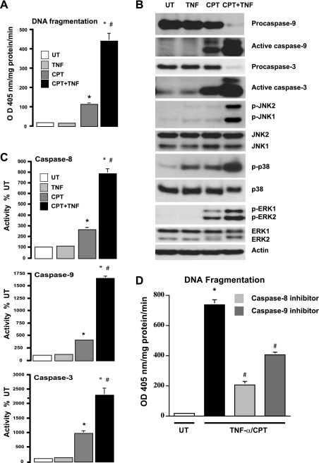
Fig. 1.
Effect of camptothecin (CPT), TNF-α, and CPT + TNF on induction of apoptosis. A: intestinal epithelial (IEC-6) cells were left untreated (UT) or exposed to TNF-α (10 ng/ml) (TNF), CPT (20 μM), or TNF-α + CPT (TNF + CPT) for 4 h. DNA fragmentation was measured using a colorimetric ELISA kit. OD, optical density. Values are means ± SE of triplicates. *Significantly different from UT. #Significantly different from CPT. B: cell extracts (25 μg) from experiments described in A were subjected to SDS-PAGE followed by Western blot analysis. Levels of procaspase-9, active caspase-9, procaspase-3, active caspase-3, and total MAPKs (ERK1/2, JNK1/2, and p38) and phosphorylated MAPKs (p-ERK1/2, p-JNK1/2, and p-p38) were determined using specific antibodies. Actin immunoblotting was performed as an internal control for equal loading. Blots are representative of 3 observations. C: confluent serum-starved cells treated as described in A were used to determine enzymatic activities of caspases-8, -9, and -3. Values are means ± SE of triplicates. *Significantly different from UT and TNF-α. #Significantly different from CPT. D: confluent serum-starved cells were treated with TNF + CPT in the presence or absence of inhibitors of caspase-8 or -9 for 4 h. Apoptosis was measured as described in A. Values are means ± SE. *Significantly different from UT. #Significantly different from CPT + TNF.

