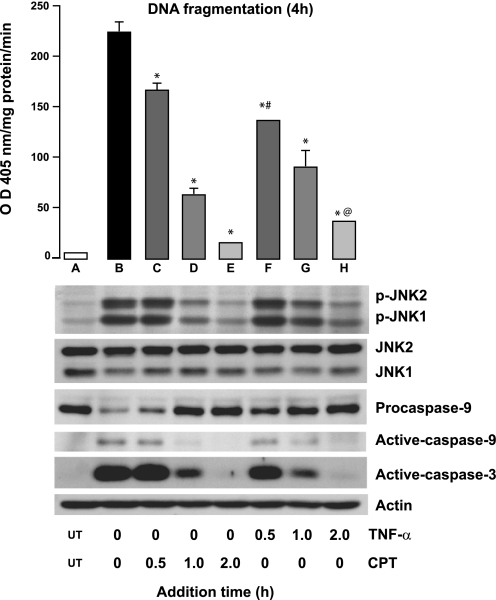
Fig. 4.
JNK1/2 activation during TNF + CPT-induced apoptosis. Confluent serum-starved cells were exposed to TNF-α (10 ng/ml) or CPT (20 μM) at 0 h, CPT and TNF-α, respectively, were added at indicated times, and cells were further incubated until 4 h. TNF + CPT at 0 h served as control. At the end of 4 h, apoptosis was evaluated by DNA fragmentation assay (top), and cell extracts were analyzed by Western blot for detection of phosphorylated JNK1/2, total JNK1/2, procaspase-9, and active caspases-9 and -3 (bottom). Values are means ± SE of 3 observations. *Significantly different from A. #Significantly different from C. @Significantly different from D.

