Fig. 2.
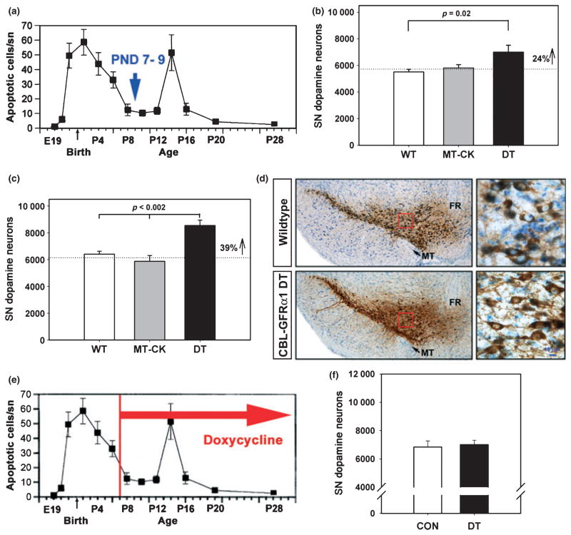
Post-synaptic GFRα1 expression in striatum increases the number of SNpc dopamine neurons that survive developmental cell death. (a) Counts of the number of apoptotic profiles in the SNpc throughout the postnatal period reveal that there are two phases of developmental cell death. The first occurs just before and after birth; the second occurs at PND14. The data shown are derived from studies of rat (data from Oo and Burke 1997); a very similar time course has been demonstrated in mice (Jackson-Lewis et al. 2000). (b) After the first phase of developmental cell death (at PND7–9), striatal GFRα1 expression results in a 24% increase in the number of dopamine neurons in the SN (p = 0.02, ANOVA; n = 7, all groups) (WT, wildtype, non-transgenic littermate control; MT-CK, monotransgenic CaMK-tTA; DT, double transgenic CBL-GFRα1 mice). (c) The increased survival of SN dopamine neurons persists through the second phase of developmental cell death and into adulthood, when a 39% increase in their number is observed (p < 0.002, ANOVA, n = 6 for the WT and MT-CK groups; n = 8 for double transgenics). For this experiment, the ages of the mice ranged from 9 to 14 weeks. (d) Coronal sections of the SN have been immunoperoxidase stained for TH (brown) and Nissl counter-stained. Low power micrographs reveal an increased number of dopamine neurons in the CBL-GFRα1 DT mice (bottom left panel). Higher magnification of the regions outlined in red squares shows that there is no apparent difference in the size or morphology of the neurons (MT, medial terminal nucleus; FR, fasciculus retroflexus) (Bar = 10 μm). (e, f) Administration of doxycycline from PND7 into adulthood suppresses the effect of post-synaptic striatal GFRα1 on the surviving adult number of SN dopamine neurons.

