
Fig. 4.
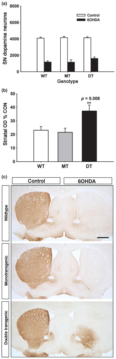
Preservation of striatal dopaminergic innervation following neurotoxin lesion by GFRα1 expression in trans. (a) Stereologic counts of dopamine neurons in the SN reveals no protective effect of striatal GFRα1 expression in the intra-striatal 6-OHDA lesion model (p > 0.05, all comparisons among 6-OHDA-treated conditions, ANOVA; n = 7, all groups). Adult (8–12 week) littermates of CaMKIIα-tTA × BiTetO-LacZ-rGFRα crosses were used, including non-transgenic (WT), monotransgenic (MT) (CaMKIIα-tTA) and double transgenic (DT) (CBL-GFRα1) genotypes. (b) In spite of the lack of protection of neurons, there is preservation of dopaminergic innervation, assessed as optical density of TH immunoreactivity. For this assessment, optical densities for the striatum on the lesioned side are normalized for the optical density of the contralateral, non-lesioned control side, and expressed as a percent of the control (p = 0.008, DT genotype vs. WT and MT, ANOVA; n = 7, all groups) (OD, optical density; CON, control). (c) Coronal sections of the striatum, immunoperoxidase stained for TH, show that the ability of GFRα1 to preserve innervation is primarily observed in the ventro-medial quadrant of the striatum. **p = 0.008.