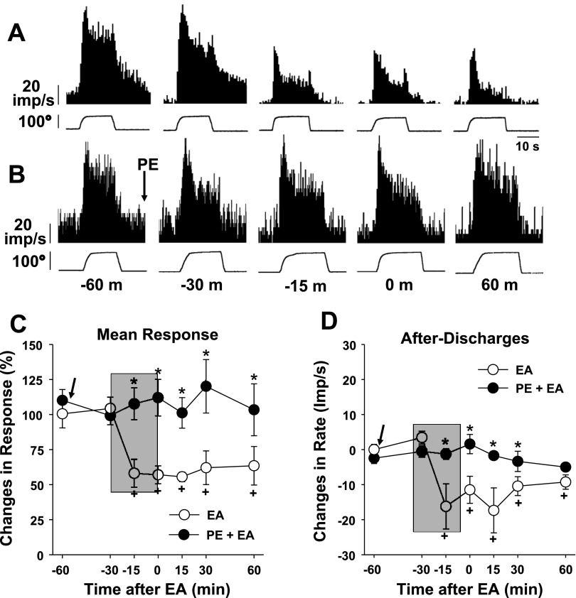
Fig. 5.
Effects of PE (5 mg/kg ip) pretreatment on the EA-induced inhibition of dorsal horn neuron activity in ankle-sprained rats. The EA period is indicated by the shaded boxes. A and B: examples of dorsal horn neuron responses to inversion of the foot before (−60 and −30 min), during (−15 min), and after (0 and 60 min) EA stimulation without (A) and with (B) PE pretreatment. C: changes of the averaged mean responses from 11 neurons (4 plantarflexion-, 3 inversion-, and 4 compression-responsive neurons combined). D: changes of averaged afterdischarges. Arrows indicate the time of PE injection on seven neurons (●) but not four neurons (○). *Values were significantly different from the control (EA) group; +values were significantly different from the pre-EA value (at −60 m).

