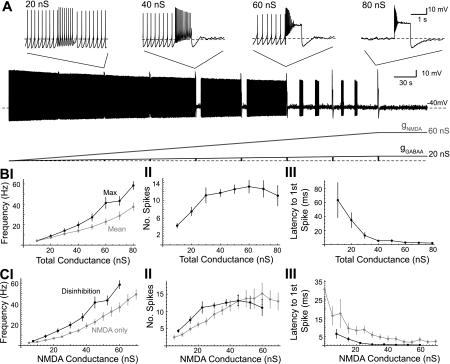
Fig. 2.
Disinhibition burst frequency increases as the total applied NMDA/GABAA conductance increases. A: concurrent NMDA and GABAA conductance ramps (3:1 ratio) were applied to a dopaminergic neuron in a representative example. As the total applied conductance reached 10, 20, ..., 80 nS, the GABAA conductance was phasically set to zero for 1 s. Insets show the change in firing at total conductance of 20, 40, 60, and 80 nS. B: summary data for the maximum and mean frequency (I), number of spikes (II), and latency to the first spike (III) during the 1-s disinhibition window as a function of the total applied conductance (n = 9). Burst onset was defined as the latency of the first spike in the first interspike interval (ISI) of <80 ms (cells in which this criteria were not met were excluded from the mean calculation for that conductance). C: maximum firing frequency (I), number of spikes (II), and latency to first spike (III) for disinhibition bursts (solid) are shown along with bursts evoked from phasic NMDA receptor activation alone (shaded). The NMDA conductance of the disinhibition burst was 0.75 times the total applied conductance. NMDA-only bursts were evoked by a 1-s NMDA conductance step (5–70 nS, 5-nS increment) in a spontaneously firing dopaminergic neuron.

