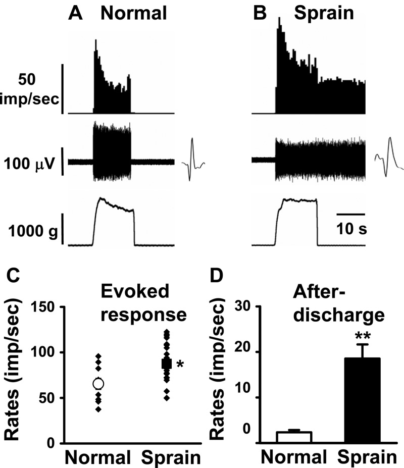
Fig. 4.
Response characteristics of spinal dorsal horn neurons to compression (Cp) of the ankle joint in normal and ankle-sprained rats. Cp was applied from the medial to lateral sides of the ankle with a pair of forceps equipped with Cp force output reading. The neurons responding exclusively to Cp were recorded in normal (A) and ankle-sprained (B) rats. Insets show raw recordings of single spikes. C: mean evoked responses to Cp. D: afterdischarges of the dorsal horn neurons recorded from normal rats (n = 10) and 1 day postankle-sprained rats (n = 20). Asterisks indicate values significantly different from the normal value (*P < 0.05; **P < 0.01).

