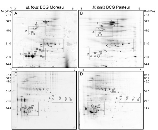
Figure 4.
Comparative 2DE profiles of CFPs from M. bovis BCG strains Moreau and Pasteur. Proteins (500 ug) were applied to IPG strips in the pH intervals of 3 - 6 (panels A and B) and 4 - 7 (panels C and D) and separated in the second dimension in 12% (panels A and B) and 15% (panels C and D) SDS-PAGE. Protein spots were visualized by colloidal CBB-G250 staining and the gels images compared with PDQuest (Bio-Rad). Molecular weight standards indicated in kDa. The sectors shown in more detail in Additional files 5 and 6, Figures S2 and S3, are indicated in the figure (sectors A - G).

