Figure 1.
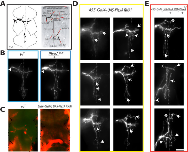
Reducing PlexinA expression increases axonal branching. A, The posterior scutellar (pSc) neuron has a stereotyped axonal branching pattern within the central nervous system. Left panel, the pSc mechanosensory neuron enters the thoracic ganglion (arrowhead) in a specific nerve bundle within the mesothoracic region. Right panel, the pSc axonal arbor consists of 16 stereotyped, "skeleton" branches (black lines), and 6 additional variable branches (not shown). Length measurements are shown in micrometers with standard deviations of individual branches indicated as red lines. B, Axon guidance into the thoracic ganglion is unaffected in PlexALOF/+ heterozygous mutants. C, PlexinA is broadly distributed throughout the thoracic ganglion. PlexinA protein expression is shown in green with MAP1B protein counterstained in red. Pan-neuronal knockdown of PlexinA using RNAi reduces PlexinA protein to undetectable levels. D, RNAi-mediated knockdown of PlexinA in the pSc axon increases the number of axonal branches (arrows). Selective expression of dsRNA constructs within pSc neurons was through the 455-Gal4 driver. E, Combining PlexinA RNAi in pSc neurons with PlexALOF mutants increases axon guidance errors (arrowheads), routing errors (asterisks), and excessive branching (arrows). Dotted lines represent the midline and the scale bar is 50 μm.

