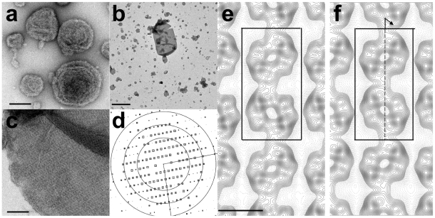
Figure 6. 2-D crystallization of BmrC/BmrD.
BmrC/BmrD was reconstituted with PC/PA/cholesterol lipid mixture at 0.6 (w/w) as described in the Materials and Methods section in the absence (a) or in the presence (b, c) of ATP/MgCl2/vanadate. (b, c) Electron micrographs of negatively stained 2-D crystals. (d) Representation of amplitudes of Fourier components calculated for one image of a negatively stained crystal. Numbers and box sizes correspond to the spot IQ value, with spots of the highest signal-to-noise ratio having an IQ of 1 and the lowest of 5 [84]. The three black circles are at radii corresponding to 1/35, 1/24, 1/18 Å−1. (e, f ) Projection maps of BmrC/BmrD at 21 Å resolution calculated from merged amplitudes and phases from five independent crystals with p1 symmetry and p121-b symmetry, respectively. Solid lines indicate density above the mean. Negative contours are indicated by dotted lines. The lattice parameters are a = 122 Å, b = 236 Å and gamma = 90°. The unit cell, dark box, contains two supramolecular entities (one being outlined by a dotted, light gray box in e), related by a screw axis along b (dotted arrow in f ). Scale bars are equal to 100 nm (a and c), 1 µm (b).

