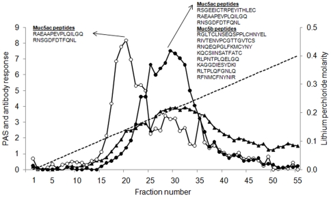
Figure 3. Separation of Muc5b and Muc5ac by anion exchange chromatography.
Purified respiratory mucins were reduced and carboxymethylated and dialysed into 6 M urea prior to anion exchange chromatography on a Resource Q column. Aliquots of each fraction were analysed by PAS-staining (black triangles) and for reactivity with MANeq5b-I (black circles) and MANeq5ac-I (open circles). The dotted line indicates the linear gradient of lithium perchlorate. Inset shows Muc5ac and Muc5b peptides identified by tandem mass spectrometry analysis of fractions 20 and 29.

