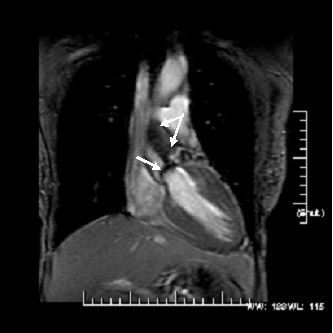
Figure 1.

A coronal view from a steady-state free precession acquisition demonstrating the heavily calcified (arrow) and restricted aortic valve leaflets with a intervoxel dephasing defect as depicted by the systolic turbulence (bifid arrow) radiating into the proximal ascending aorta. In itself, this is indictative of a highly velocity jet consistant with severe AS. Using phase velocity mapping to formally quantitate the mean and peak transvalvular gradients, they were 53 and 78 mmHg, respectively; severe AS.
