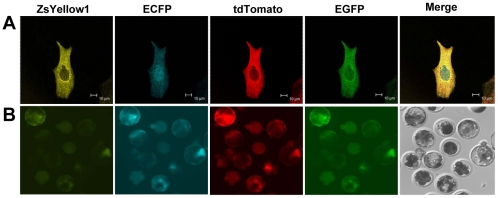
Figure 2. Co-expression of the four fluorescent proteins in PFFs and blastocysts.
(A) PFFs transfected with the linearized pZCpTG vector were observed under a confocal microscope using appropriate filters. The scale bar represents 10 µm. (B) Several reconstructed embryos derived from the pZCpTG PFFs were cultured until they reached the blastocyst stage. The blastocysts were observed under a fluorescence microscope using appropriate filters.

