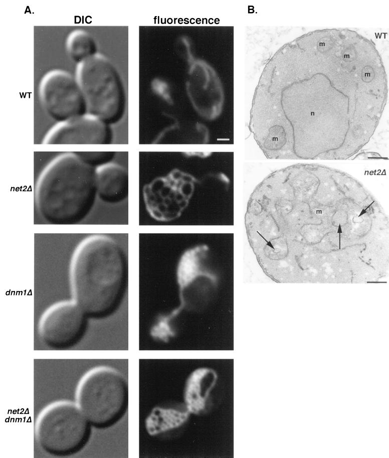
Figure 2.
net2Δ cells contain a single mitochondrion composed of an interconnected network of tubules. (A) Wild-type strain BY4742 (WT), net2Δ strain RJ1253, dnm1Δ strain RJ1188, and net2Δ dnm1Δ strain 1285, each expressing matrix-targeted GFP from pHS12 (Sesaki and Jensen, 1999), were grown to mid log phase in synthetic medium with 2% galactose and examined by DIC and fluorescence microscopy. Representative images of cells are shown. Bar, 5 μm. (B) Wild-type and net2Δ cells were fixed, embedded, sectioned, and examined by electron microscopy. Arrows indicate spaces that result from the interconnected network of tubules that forms a single mitochondrion in the net2Δ cells. m, mitochondria; n, nucleus. Bar, 0.1 μm.

