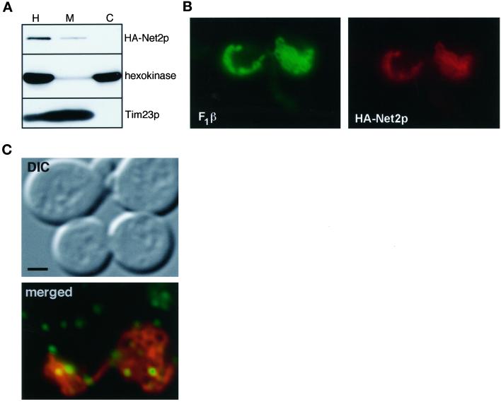
Figure 8.
Net2p and Dnm1p do not recruit each other to the mitochondrial outer membrane. (A) Location of Net2p in dnm1Δ cells. dnm1Δ cells, strain RJ1188, expressing HA-Net2p from pKC5 were grown to OD600 of 0.8 in synthetic medium with 2% galactose and subjected to cell fractionation. Aliquots of homogenate (H), cytosol (C), and mitochondria (M) representing equivalent numbers of cells were subjected to SDS-PAGE and analyzed by Western blotting with antibodies against the HA epitope (HA-Net2p), hexokinase, and Tim23p. (B) Formation of punctate structures containing Net2p requires Dnm1p. net2Δ dnm1Δ cells (RJ1285), expressing HA-Net2p from pKC5, were fixed, permeabilzed, and then incubated with antibodies to the HA epitope and the mitochondrial F1β protein. Immune complexes were visualized with FITC-conjugated antibodies (F1β) and CY3-linked antibodies (HA-Net2p). Representative images from the green (F1β) and red (HA-Net2p) channels are shown. (C) Localization of Dnm1p in net2Δ cells. net2Δ strain RJ1253, constitutively expressing Dnm1p-GFP from pHS20 (Sesaki and Jensen, 1999), was transformed with pHS51, which expresses Cox4-RFP under the control of the galactose-inducible GAL1 promoter. Cells were grown in media with 2% raffinose to an OD600 of 0.3 and then resuspended in synthetic medium containing 2% galactose and 2% sucrose for 2.5 h to induce expression of matrix-targeted RFP. Cells were then examined by DIC and fluorescence microscopy. A representative merged image from the red (cox4-RFP mitochondria) and green (Dnm1p-GFP) is shown. Bar, 5 μm. Note that the upper two cells are not expressing Cox4-RFP.

