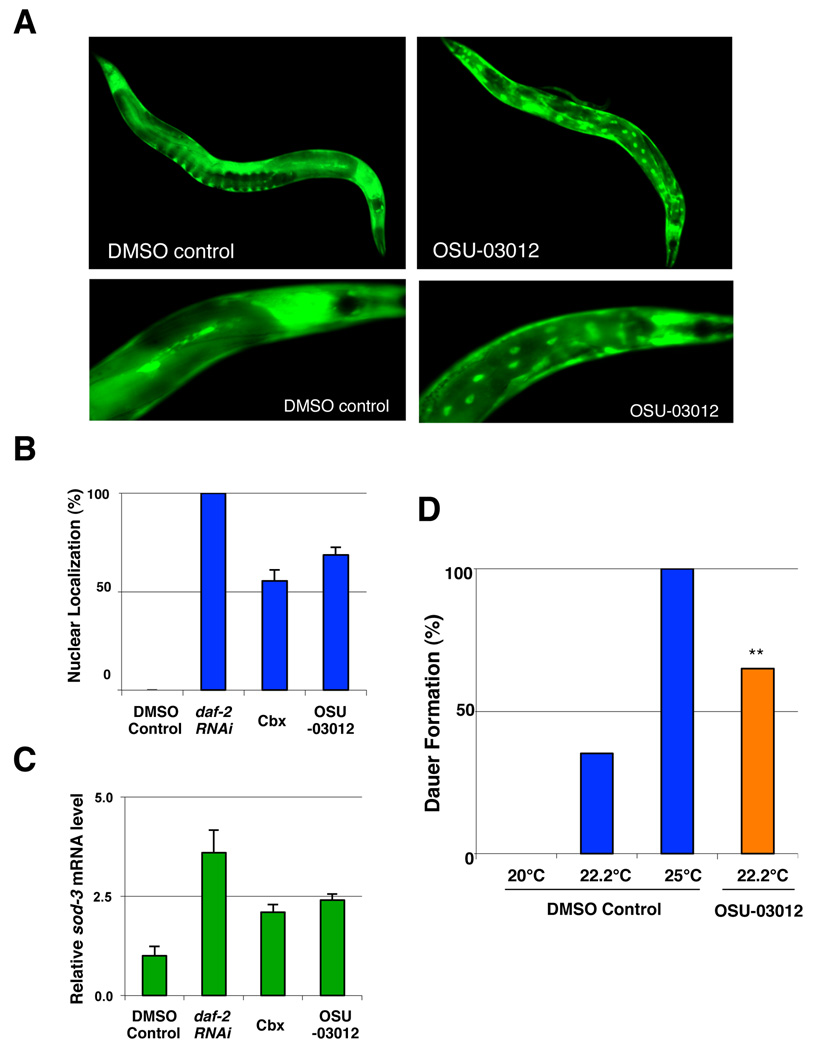
Figure 6. OSU-03012 alters the nuclear localization and the activity of DAF-16.
(A) Nuclear accumulation of DAF-16 induced by OSU-03012 treatment. Images of TJ356 animals, which carry an integrated daf-16::gfp array in a wild-type background, exposed to either DMSO control or 0.5 µM of OSU-03012 for 72 hours. (B) Quantification of the nuclear accumulation of DAF-16::GFP in response to 10 µM celecoxib and 0.5 µM OSU-03012 treatments. Worms were scored for the presence or absence of GFP accumulation within the intestinal nuclei at the first day of adulthood (n ≥ 120 for each treatment). An animal was scored as having nuclear DAF-16 if more than one intestinal nucleus contained DAF-16-GFP. RNAi treatment by feeding of daf-2 was also performed as a positive control. Lifespans following each drug treatment were determined to confirm the effectiveness of the drug treatment. (C) Effects of celecoxib and OSU-03012 on DAF-16 transcriptional activity. Wild-type N2 animals were exposed to DMSO control, 10 µM celecoxib, or 0.5 µM OSU-03012 from hatching. Relative mRNA levels of sod-3 of these animals were measured by quantitative RT-PCR and the mean of three different sample sets are shown. The relative mRNA levels were normalized against act-1 (beta-actin) levels. Error bars: ± STD. (D) daf-2(e1370) mutants (P0) were exposed to DMSO control or 0.5 µM OSU-03012 at 20°C. The F1 eggs were then moved to the different temperatures indicated for 72 hours before being scored for dauer formation. Each bar represents combined data of three independent experiments per condition (n ≥ 150 for each treatment). Asterisks indicate significant changes (*, P < 0.005). P values were calculated by Pearson's chi-square test.

