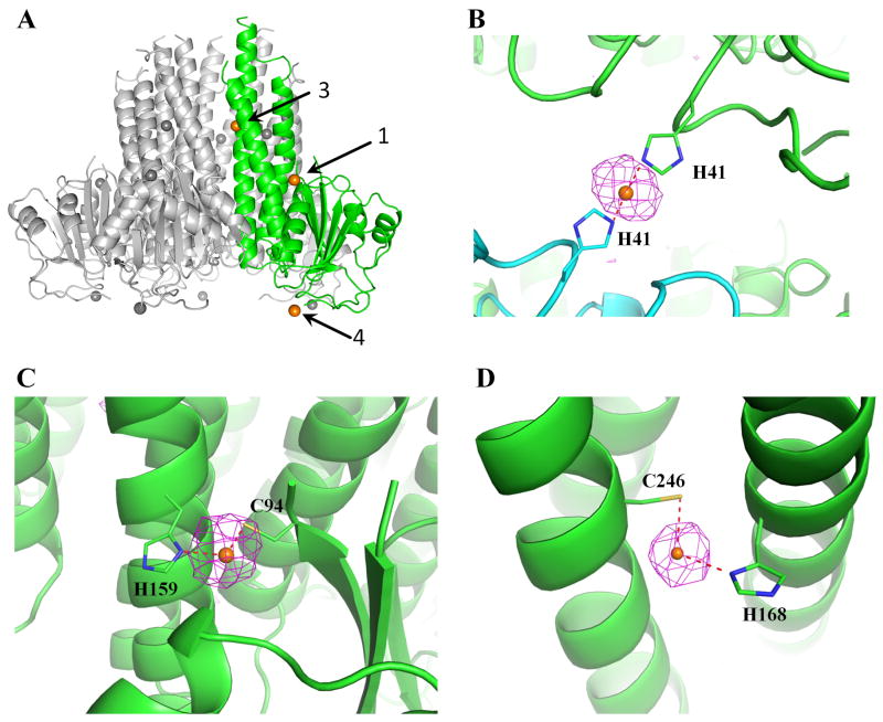
Figure 3. Zn2+ binding sites in Salmonella ZntB.
(A) The three Zn2+ (orange) binding sites in the monomer (green) within the soluble domain pentamer are labeled 1, 3 and 4. Other monomers are in gray. (B) The anomalous difference map contoured at 3σ is shown for site 4, where Zn2+ is coordinated by two neighboring His41 residues related by crystal packing. (C) Site 1 on the outside surface of the funnel where Zn2+ is coordinated by C94 and H159 of the same monomer and (D) in site 3 Zn2+ is bound inside the interior wall of the funnel by H168 and C246 of the same monomer. See also Supplementary Figure 3.

