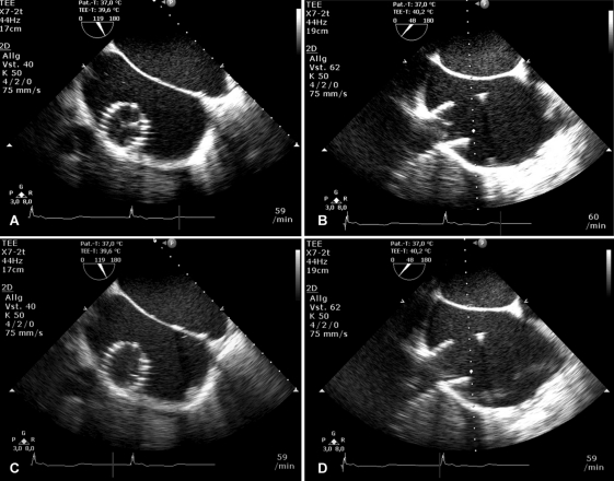
Figure 3.
Echocardiographic evaluation of prosthetic valve function systolic closure (A and B) and diastolic opening (C and D) of the pericardial valve is visualized by transoesophageal echocardiography after the implantation procedure. Short- and long-axis views of the device after deployment. Doppler interrogation confirmed valvular competence and a continuously antegrade blood flow in the inferior vena cava and the hepatic veins without flow reversal.

