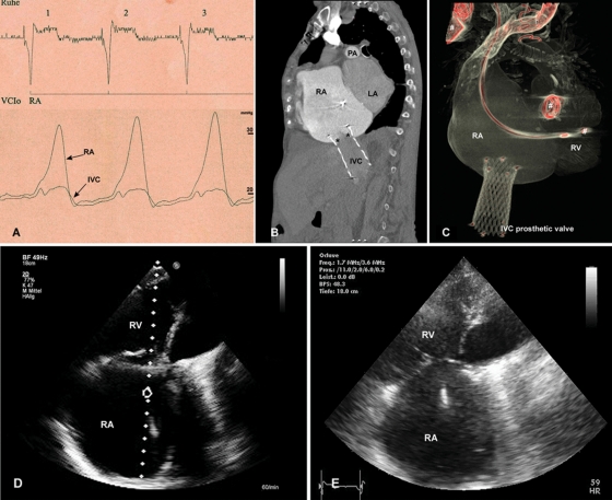
Figure 4.
After device implantation, (A) deployment of the self-expanding valve resulted in the immediate abolition of regurgitation into the inferior vena cava. Simultaneous pressure measurement in the right atrium and the inferior vena cava confirms a reduction in the v-wave. (B) A computed tomographic angiogram 8 weeks after implantation with contrast injected in superior vena cava visualizes valve function without backflow into the inferior vena cava. The level of the leaflets (asterisks) is aligned with the cavoatrial junction, thus protecting of the hepatic vein from elevated right atrium pressure. (C) Three-dimensional reconstruction confirms the forward-tilted position of the valve in the inferior vena cava. (D) Transthoracic echo after 8 weeks visualizes right atrial and right ventricular enlargement; however, right heart morphology and function remained unchanged compared with the (E) pre-operative state. RA, right atrium; RV, right ventricle; IVC, inferior vena cava; LA, left atrium; PA, pulmonary artery. Previously implanted devices (pacer leads, prosthetic mitral valve) are marked with #.

