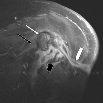
Fig. 3.
A coronal postcontrast T1-weighted MR image shows peripheral rim enhancement of the fluid collection (thick black arrow) and an enhancing fistulous tract leading to the skin surface (white block arrow). There is mild enhancement of the residual humeral head (thin white arrow) without appreciable enhancement of the glenoid (thin black arrow.)

